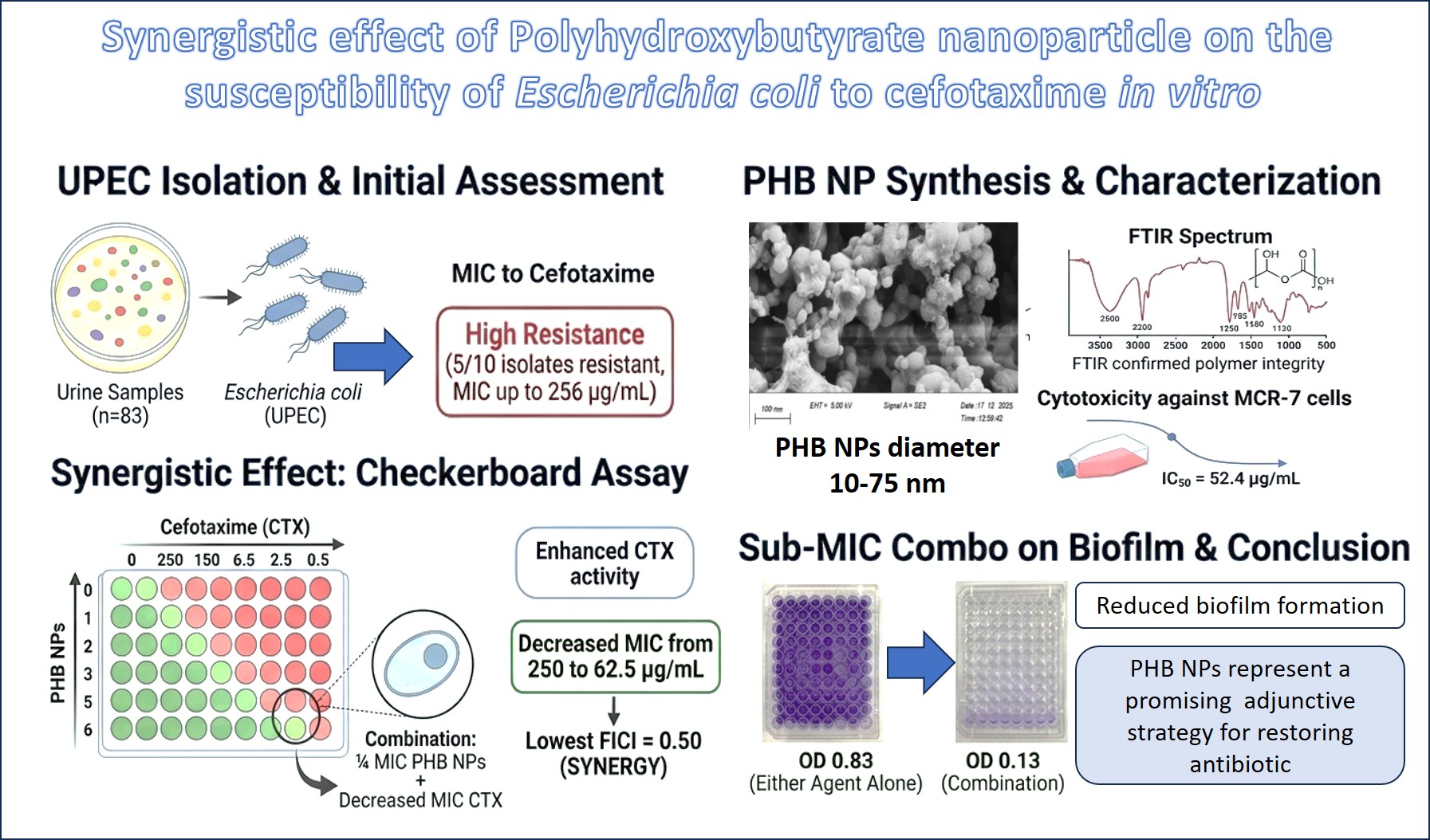
Synergistic effect of Polyhydroxybutyrate nanoparticle on the susceptibility of Escherichia coli to cefotaxime in vitro
PHB Nanoparticles Enhance Cefotaxime Activity Against E. coli
DOI:
https://doi.org/10.65329/wjeb.v14.01.07Keywords:
Antimicrobial resistance; Biofilm eradication; Biopolymer nanoparticles; Cefotaxime; Drug-resistant; Green nanotechnology; Nanomedicine; PHP-NPs; Uropathogenic E. coli.Abstract
Multidrug-resistant uropathogenic Escherichia coli poses a major therapeutic challenge. Nanotechnology is an alternative strategy to improve the effectiveness of conventional antibiotics. The synergistic effect of nanoparticles in restoring antibiotic susceptibility is scarce in the literature. The study aims to evaluate the synergistic effect of polyhydroxybutyrate (PHB) nanoparticles on UPEC susceptibility to cefotaxime (CTX) using a checkerboard assay. The effect of the combination on UPEC's ability to form biofilms was evaluated. The UPEC was isolated from 83 urine samples. The most isolates were resistant to CTX (5/10), and the minimum inhibitory concentration (MIC) ranged from 0.062 to 256 µg/mL. Biofilm formation showed a significant correlation with resistance to CTX. The PHB NPs were synthesized and characterized by scanning electron microscopy, which revealed a diameter of 10-75 nm, and by FTIR spectroscopy, which confirmed polymer integrity. The cytotoxicity assessment against MCR-7 cells yielded an IC50 of 52.4 µg/mL. Checkboard microdilution assays against the resistant and strong biofilm-form isolates of E. coli (Ec9) showed that PHB NPs enhanced CTX activity, decreasing its MIC from 250 µg/mL to 62.5 µg/m. The combination of ¼ MIC PHB NPs produced the lowest fractional inhibitory concentration (FICI; 0.50), indicating a synergistic interaction. The sub-MIC combination of the two agents significantly reduced biofilm formation compared to either agent alone, resulting in an optical density (OD) decrease from 0.83 to 0.13. From the current study, it can be concluded that PHB NPs represent a promising adjunctive strategy for restoring antibiotic activity. These results need further research to achieve the final goal of reactivating conventional antibiotics.
References
[1] Ranjbar R, Alam M. (2023) Antimicrobial Resistance Collaborators (2022). Global burden of bacterial antimicrobial resistance in 2019: a systematic analysis. Evid Based nurs ebnurs-2022-103540. doi: https:doi.org/10.1136/ebnurs-2022-103540 . PMID: 37500506 DOI: https://doi.org/10.1136/ebnurs-2022-103540
[2] Bonten M, Johnson JR, van den Biggelaar AHJ, Georgalis L, Geurtsen J, et al. (2021) Epidemiology of Escherichia coli Bacteremia: A Systematic Literature Review. Clin Infect Dis 72(7): 1211–1219. doi: https:doi.org/10.1093/cid/ciaa210 , PMID: 32406495. DOI: https://doi.org/10.1093/cid/ciaa210
[3] Nasrollahian S, Graham JP, Halaji M. (2024) A review of the mechanisms that confer antibiotic resistance in pathotypes of E. coli. Front Cell Infect Microbiol 14: 1387497. doi: https:doi.org/10.3389/fcimb.2024.1387497 . PMCID: PMC11024256. DOI: https://doi.org/10.3389/fcimb.2024.1387497
[4] Wang R, Degnan KO, Luther VP, Szymczak JE, Goren EN, et al. (2021) Development of a Multifaceted Antimicrobial Stewardship Curriculum for Undergraduate Medical Education: The Antibiotic Stewardship, Safety, Utilization, Resistance, and Evaluation (ASSURE) Elective. Open Forum Infect Dis 8(6): ofab231. doi: https:doi.org/10.1093/ofid/ofab231. PMCID: PMC8215691 DOI: https://doi.org/10.1093/ofid/ofab231
[5] Rufino AT, Lucas M, Silva AMS, Ribeiro D, Fernandes E. (2023) 2-Styrylchromones Prevent IL-1β-Induced Pro-Inflammatory Activation of Fibroblast-like Synoviocytes while Increasing COX-2 Expression. Pharmaceutics 15(3): 780. doi: https:doi.org/10.3390/pharmaceutics15030780. PMCID: PMC10053337 . DOI: https://doi.org/10.3390/pharmaceutics15030780
[6] Modi SK, Gaur S, Sengupta M, Singh MS. (2023) Mechanistic insights into nanoparticle surface-bacterial membrane interactions in overcoming antibiotic resistance. Front Microbiol 14: 1135579. doi: https:doi.org/10.3389/fmicb.2023.1135579. PMCID: PMC10160668 DOI: https://doi.org/10.3389/fmicb.2023.1135579
[7] Sadiq SI, Ghafil JA. (2025) Polyhydroxybutyrate nanoparticle improving the sensitivity of Pseudomonas aeruginosa to ceftriaxone and reducing the biofilm formation in vitro. Polim Med 55(1): 31–37. doi: https:doi.org/10.17219/pim/203765 . PMID: 40599100 DOI: https://doi.org/10.17219/pim/203765
[8] Yang JW, Shen YC, Lin KC, Cheng SJ, Chen SL, et al. (2020) Organ-on-a-Chip: Opportunities for Assessing the Toxicity of Particulate Matter. Front Bioeng Biotechnol 8: 519. doi: https:doi.org/10.3389/fbioe.2020.00519. PMCID: PMC7272695. DOI: https://doi.org/10.3389/fbioe.2020.00519
[9] Talib MM, Ghafil JA. (2024) Comparative Adhesion of Pseudomonas aeruginosa to Human Oral Mucosal Epithelial Cells and Polystyrene Surfaces. J Faculty Med Baghdad 66(3): 344-349. doi: https:doi.org/10.32007/jfacmedbaghdad.6632328. DOI: https://doi.org/10.32007/jfacmedbaghdad.6632328
[10] Clinical and Laboratory Standards Institute. (2024) Performance standards for antimicrobial susceptibility testing; 34th edition. CLSI supplement M100. Clinical and Laboratory Standards Institute.
[11] Mosmann T. (1983) Rapid colorimetric assay for cellular growth and survival: application to proliferation and cytotoxicity assays. J Immunol Methods 65(1-2):55-63. doi: https:doi.org/10.1016/0022-1759(83)90303-4. DOI: https://doi.org/10.1016/0022-1759(83)90303-4
[12] Plumb JA. (1999) Cell sensitivity assays: the MTT assay. Methods Mol Med 28:25-30. doi: https:doi.org/10.1385/1-59259-687-8:25. DOI: https://doi.org/10.1385/1-59259-687-8:25
[13] Berridge MV, Herst PM, Tan AS. (2005) Tetrazolium dyes as tools in cell biology: new insights into their cellular reduction. Biotechnol Annu Rev 11:127-52. doi: https:doi.org/10.1016/S1387-2656(05)11004-7. DOI: https://doi.org/10.1016/S1387-2656(05)11004-7
[14] Al-Mutalib LAA, Zgair AK. (2023) Effect of subinhibitory doses of rifaximin on in vitro Pseudomonas aeruginosa adherence and biofilm formation to biotic and abiotic surface models. Polim Med 2023;53(2):97-103. doi: https:doi.org/10.17219/pim/166584. DOI: https://doi.org/10.17219/pim/166584
[15] Talib MM, Ghafil JA. (2024) Effect of sub-minimum inhibitory concentrations of ceftriaxone on the Pseudomonas aeruginosa adhesion to human oral mucosal epithelial cells and biofilm formation to polystyrene in vitro. Pharm Sci Asia 51:180-189. doi: https:doi.org/10.29090/psa.2024.02.24.1752. DOI: https://doi.org/10.29090/psa.2024.02.24.1752
[16] Ibrahim B, Ghafil JA, Abdullah ZA, Kınaytürk NK, Alshahrani SM, Khan BA, Zgair AK. (2026) Molecular insights into the oxidative perturbation of VIM-2 metallo-β-lactamase: Active site remodeling restores imipenem susceptibility in Pseudomonas aeruginosa. Microb Pathog 214: 108411. doi: https:doi.org/10.1016/j.micpath.2026.108411 DOI: https://doi.org/10.1016/j.micpath.2026.108411
[17] Sati H, Carrara E, Savoldi A, Hansen P, Garlasco J, et al. (2025). The WHO Bacterial Priority Pathogens List 2024: a prioritisation study to guide research, development, and public health strategies against antimicrobial resistance. Lancet Infect Dis 25(9): 1033–1043. doi: https:doi.org/10.1016/S1473-3099(25)00118-5. PMCID: PMC12367593. DOI: https://doi.org/10.1016/S1473-3099(25)00118-5
[18] Mouhammed K, Gdoura R. (2024) Study of the Genomic Characterization of Antibiotic-Resistant Escherichia coli Isolated From Iraqi Patients with Urinary Tract Infections. Indian J Microbiol 64(2): 457–466. doi: https:doi.org/10.1007/s12088-023-01123-3. PMCID: PMC11246310 DOI: https://doi.org/10.1007/s12088-023-01123-3
[19] Alshaikh SA, El-Banna T, Sonbol F, Farghali MH. (2024) Correlation between antimicrobial resistance, biofilm formation, and virulence determinants in uropathogenic Escherichia coli from Egyptian hospital. Ann Clin Microbiol Antimicrob 23(1): 20. doi: https:doi.org/10.1186/s12941-024-00679-2. PMCID: PMC10894499 DOI: https://doi.org/10.1186/s12941-024-00679-2
[20] Usui M, Yoshii Y, Thiriet-Rupert S, Ghigo JM, Beloin C. (2023) Intermittent antibiotic treatment of bacterial biofilms favors the rapid evolution of resistance. Commun Biol 6(1): 275. doi: https:doi.org/10.1038/s42003-023-04601-y. PMCID: PMC10020551. DOI: https://doi.org/10.1038/s42003-023-04601-y
[21] Manal Munir F, Safdar W, Abu Bakr Shabbir M, Ahmed S, Navid MT, et al. (2025) Production, characterization, and antimicrobial activity of polyhydroxyalkanoates synthesized by Bacillus species against skin pathogens. RSC adv 15(42): 35182–35200. doi: https:doi.org/10.1039/d5ra04375a. PMCID: PMC12459338 DOI: https://doi.org/10.1039/D5RA04375A
[22] Campos JV, Pontes JTC, Canales CSC, Roque-Borda CA, Pavan FR. (2025) Advancing Nanotechnology: Targeting Biofilm-Forming Bacteria with Antimicrobial Peptides. BME front 6: 0104. doi: https:doi.org/10.34133/bmef.0104. PMCID: PMC1187654 DOI: https://doi.org/10.34133/bmef.0104
[23] Lu L, Zhao Y, Li M, Wang X, Zhu J, et al. (2024) Contemporary strategies and approaches for characterizing composition and enhancing biofilm penetration targeting bacterial extracellular polymeric substances. J Pharm Anal 14(4): 100906. doi: https:doi.org/10.1016/j.jpha.2023.11.013. DOI: https://doi.org/10.1016/j.jpha.2023.11.013
[24] Mishra S, Gupta A, Upadhye V, Singh SC, Sinha RP, Häder DP. (2023) Therapeutic Strategies against Biofilm Infections. Life (Basel): 13(1): 172. doi: https:doi.org/10.3390/life13010172. PMCID: PMC9866932 DOI: https://doi.org/10.3390/life13010172
[25] Gupta A, Makabenta JMV, Schlüter F, Landis RF, Das R, et al. (2020) Functionalized Polymers Enhance Permeability of Antibiotics in Gram-negative MDR Bacteria and Biofilms for Synergistic Antimicrobial Therapy. Adv ther 3(7): 2000005. doi: https:doi.org/10.1002/adtp.202000005. PMCID: PMC907568. DOI: https://doi.org/10.1002/adtp.202000005
[26] Afrasiabi S, Partoazar A. (2024) Targeting bacterial biofilm-related genes with nanoparticle-based strategies. Front Microbiol 15: 1387114. doi: https:doi.org/10.3389/fmicb.2024.1387114. PMCID: PMC11150612 . DOI: https://doi.org/10.3389/fmicb.2024.1387114
Downloads
Additional Files
Published
Issue
Section
License
Copyright (c) 2026 Authors

This work is licensed under a Creative Commons Attribution 4.0 International License.
All articles in the World Journal of Experimental Biosciences are published under the terms of the Creative Commons Attribution 4.0 International License (CC BY 4.0) ( (http://creativecommons.org/licenses/by/4.0/), which permits unrestricted use, distribution, and reproduction in any medium, provided the original work is properly cited.